This lesson will provide an overview to maxillofacial trauma, and we will explore some of the principles behind its clinical features and management. Fasten your seatbelt, and get ready for loads of diagrams and new terminology that will come in the next few lessons!
From high impact road traffic accidents to violence, or maybe just a "simple" fall; facial trauma is still a common injury that presents in our accident and emergency departments, and may present first in the dental chair. We must be able to recognised these injuries to make appropriate referrals, gain an understanding of their management and any complications that can occur, ready for when the patient is discharged back into primary care.
Yes, and there are four key ways in which facial fractures differ from fractures of bone in other parts of the body:
Following a facial bone fracture, the affected bone may displaced in a direction which may compromise the airway. This happens commonly with mandibular fractures, where the tension in the muscles will pull the bone fragments, along with the tongue, posteriorly towards the airway. In maxillary fractures, the maxilla may fall inferiorly due to the fracture, and thus the airway is occluded. Avulsed teeth and/or fragments of teeth may also obstruct the airway. In addition, the loss of muscle tone following injury can lead to risks to the airway (1). Regardless of the cause, any respiratory obstruction caused by a facial fracture must be managed immediately, as a compromised airway could be fatal.
These types of injuries can be associated with maxillofacial trauma (2), as the forces exerted on the facial bones can be transmitted through to the cervical spine. More importantly, cervical spine injuries must be ruled out when any injuries to the head occur, due to their high potential for mortality and neurologic morbidity (3).
You don’t find teeth on any other bones in the body! As we mentioned in the previous lesson, bones need to be brought into close apposition, to allow for primary healing to take place. Teeth can help in identifying the exact position in which the fragments need to be placed, allowing the fractured bone to retain the correct anatomical shape after healing. Furthermore, the presence of teeth on the maxilla and mandible will help with achieving stabilisation of the fracture. This means, that not only will the bones stay in place, the occlusion can be checked as the fractured bone is stabilised.
On the flip-side, teeth involved with the fracture may need to be extracted, for example: if they are infected (apical periodontitis), as there is a risk that the infection in the tooth may induce an infection in the fracture. This will be covered later in this lesson.
The facial bones have an excellent blood supply, and going through bone healing in the previous lesson should have highlighted how important this is. The facial bones have a proportionally large amount of cancellous bone. It is this cancellous bone which houses a complex network of blood vessels, where the vasculature is intertwined through many connections, called ‘anastomoses’. This system of anastomosing vessels is designed to deliver a rich blood supply, and so if there is more cancellous bone (which is highly vascular), the blood supply is vastly improved. It should be noted that the mandible has more compact bone and less cancellous bone when compared to other facial bones, thus its blood supply is reduced.
General presenting features of these types of injuries include pain, swelling, loss of function, malocclusion and/or altered sensation. This means that patients who have experienced some form of maxillofacial trauma may be driven by any of these symptoms to be seen.
Furthermore, an incident of maxillofacial trauma may be accompanied by life threatening injuries to the head and/or cervical spine (4), which can impact on the patient’s level of consciousness. A study has shown that patients suffering from maxillofacial fractures are more likely to experience intracranial haemorrhage than those who have not (5).
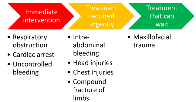
Another study demonstrated that nearly one-in-five patients with maxillofacial trauma had experienced a closed head injury (6). These types of injuries must be ruled out during the assessment phase of management, to ensure that they are not missed when delivering immediate treatment. In addition to this, there may be other pressing conditions that must be examined urgently, which will be covered now:
When a patient attends the A&E department in the hospital, the preliminary treatment that the team carries out is vital to the success of subsequent treatment of the injury. More importantly, the wellbeing of the injured patient is improved. Patients with maxillofacial trauma may present in a critical condition; therefore, it is imperative to follow a strict system to ensure that everything is stabilised. The Advanced Trauma and Life Support (ATLS) algorithm will often be adopted when assessing patients with life threatening conditions.
This system involves assessing five areas; airway, breathing, circulation and haemorrhage control, dysfunction of the central nervous system and exposure. Along with the ABCDE approach, vitals such as respiratory and heart rates, and blood pressure, all must be monitored and recorded regularly. The National Early Warning Score (NEWS) chart system is designed to help identify any deterioration in vitals and dictate what is needed next for the patient to be stabilised. In addition to monitoring vitals, the assessment of the patient's alertness is measured: Alert, Voice (respond to command), Pain (respond to painful stimulus), Unresponsive (AVPU) Note: you don't want to be in the PU.
Earlier in this lesson, we gave some examples as to why maxillofacial trauma can lead to risks to the airway. The first key thing to note is that a patent AIRWAY is established and maintained, and the lungs must be well-ventilated. Maxillofacial injuries will often involve bones which contribute towards the oral cavity, and so if they are subject to trauma, the airway may become compromised.
Following on from this, BREATHING must be maintained too, as this can also be complicated by maxillofacial trauma.
Apart from the airway being compromised from such injuries, patients presenting with maxillofacial trauma may have severe injuries elsewhere in the body too, which may lead to HAEMORRHAGE. Haemorrhage rarely occurs solely due to maxillofacial trauma, but massive bleeding will require immediate intervention if it is present. Risk of acute haemorrhage is more likely to occur if the maxillary artery or pterygoid venous plexus of the facial region and the carotid a. and jugular v. in the region of the neck.
Bleeding can be managed immediately through the use of pressure dressings and wound packing. Clamping the ruptured blood vessels will also assist in controlling the haemorrhage.
This area of the assessment focuses on the DISABILITY (medical history) and neurological status of the patient. A systematic approach through the use of the Glasgow COMA scale (GCS) helps to score the patient’s consciousness levels, which in turn will dictate the next steps in the management of the patient. The cervical spine would also be assessed by palpation and radiographic series.
A complete examination of the patient, done by EXPOSING the skin of the patient. This may help reveal more signs to build the picture and show the extent of the injuries due to the accident.
Other areas which are covered in the immediate management include shock management and resuscitation, if needed. Antibiotic prophylaxis to prevent infection of the fracture site and pain relief can also be provided. It is vital to remember that the examination and treatment of the maxillofacial fracture will often take place after more pressing issues such as respiratory obstruction and cardiac arrest. Temporary immobilisation of the suspected fracture can be considered only if it is required and once everything else has been managed appropriately.
A complete history of the injury is crucial. Important questions need to be answered here; however, in some cases the patient may not have the capacity to answer, due to an impairment in consciousness. In this case, witnesses or those accompanying the patient will need to help build the picture.
The what, when, where, who, how...
Examination of the fracture should only be conducted once the cardiopulmonary and neurological status has been assessed. If this has been carried out and all is deemed satisfactory, then a more detailed examination of the facial bones, using a systematic approach can occur.
Note: below is a generalised list and so to avoid repetition, site specific examinations will be covered in more detail in their appropriate lessons.
Following the extra-oral examination of the facial bones and structures, an intra-oral assessment is also required:
Note: It can also be useful to record findings with medical photographs for all of the above (medicolegal considerations needed - consent).
life treating injuries should be prioritised first and followed by sight threatening injuries. Unstable mandibular fractures alongside lacerations should ideally be dealt with within the first 24hrs of hospital admission. Other facial injuries may be delayed and dependent on the amount of oedema present, note: the surgeon may be able to operate before the oedema is too great. As mentioned a full body assessment may be necessary and any major chest, abdomen, pelvic or limb trauma may need to be prioritised and also delay some of the facial surgery.
Imaging will be covered in more detail in the injury specific lessons. Common images taken to assess facial trauma are:
GDPs may not necessarily treat the injury, they do; however, have adequate training in carrying out a thorough exam before referral. This will include taking a history, assessing (as best as possible) the clinical presentation, and processing an appropriate referral for further investigations and treatment through secondary care. Patients with suspected facial fractures should be direct to the closest A&E unit, and contact should be made with the on-call maxillofacial senior house officer in advance (9).
Complications can occur due to facial fractures, as well as their management. Some will happen earlier in the patient journey than others, but you may come across some of these complications in general practice, so it’s worth knowing why they occur and what can be done about them.
As mentioned previously, there is the risk of a compromised airway, and causes can pre-operative and/or post-operative. Causes for an airway obstruction include the tongue collapsing posteriorly, posterior mandibular displacement, inferior maxillary displacement, trismus, and swelling of the floor of mouth. These can create difficulties in maintaining a patent airway prior to treatment.
These complications can also occur after treatment, hence the use of drains. The patient aspirating loose debris, for example: pieces of tooth, can also compromise the airway; again emphasising on the importance of immediate airway management.
Management of a compromised airway can include correct patient positioning, chin-lift manoeuvre, and airway adjuncts such as nasopharyngeal and oropharyngeal tubes(10). Often, the mandible can be manipulated into a more anterior position to allow the tongue to move into a more favourable position. A cricothyroidotomy would be justified for airways which are completely blocked.
Fractures of the zygomatic complex and/or orbit are likely to cause compression of the infra-orbital nerve. If this occurs, anaesthesia of the nerve will take place, leading to numbness in the ipsilateral skin of the cheek, lateral nose and upper lip. Mandibular fractures can lead to damage to the inferior alveolar nerve. In both cases, damage could be permanent, and if any resolution was to occur it could take months.
The fracture site may become infected due to various reasons, and these can be grouped into trauma- and patient-related risk factors (11). Note:fractures could become infected pre-operatively and/ or post-operatively. Trauma-related risk factors for infection of a fracture include:
Broad-spectrum antibiotic prophylaxis may be indicated. This would be provided to the patient in the hospital setting if deemed appropriate. An emphasis must be on the high level of debridement in the prevention and treatment of infection.
Adverse healing complications often become noticed later, compared to the aforementioned complications. There are local and general factors which may delay healing. Local factors include:
Systemic factors include old-age, conditions such as diabetes, medications such as steroids and malnutrition. If post-operative instructions are not followed by the patient, poor healing and other complications are more likely to occur.
If there are any issues with healing, remembering that there are both local and systemic factors will help with decisions on what needs addressing first. The patient may also need multidisciplinary care in order to cope with the potential psychological effects of the trauma, especially if deformity is unavoidable.
Hopefully this lesson has given you an overview of maxillofacial trauma, and you should now have an understanding of the presentation, management and complications of facial fractures. Specific injuries will now be looked at in more detail in the coming lessons.
General references
Dimitroulis G, Avery BS. Maxillofacial Injuries: a synopsis of basic principles, diagnosis and management. Wright; 1994
Pigadas N, Oliver R. Common facial fractures: 1. Aetiology and Presentation. Dental Update 2006; 33: 347-356
Pigadas N, Oliver R. Common facial fractures: 2. Management. Dental Update 2006; 33: 413-420
Pigadas N, Oliver R. Common facial fractures: 3. Complications. Dental Update 2006; 33: 470-476
Hupp JR, Ellis III E, Tucker MR. 2014. Contemporary Oral and Maxillofacial Surgery. 6th ed. Elsevier
In-text references