The formation of the facial structures is a series of proliferations, invaginations and folds controlled by cell to cell communications. Having a fundamental understanding of these processes allows us to understand their clinical relevance when things go wrong for example, clefts. Thank you to @mydentalnotebook for the wonderful images.
The process below, involve growth, morphogenesis, differentiation, and pattern formation, controlled by gene expression and cell interaction.
Week 4:
Pharyngeal arches formed
Tongue development
Week 5:
Facial Prominences and Placodes: olfactory (nasal), optic (lens) and otic present
Week 6:
Naso-optic furrows and nasal lacrimal duct development.
Palatal shelves formation.
Week 8:
Elevation of the Palatine processes
Week 10- 12:
Fusion completion of the palate and facial processes
Week 12-16:
Bone formation of the facial structures
Soft palate formation
Note: the dates should be considered as an average, and a range can be seen dependent of the resource provided.
The stomodeum (primitive oral cavity) is evident which is initially separated from the developing pharynx, by the oropharyngeal membrane in the first 4 weeks of development.
Ectodermal thickenings form the nasal placodes. Growth of the frontonasal process alongside the invagination of the placodes occurs to form the nasal pits. The tissue either side of the pits are now clearly the medial and lateral nasal processes. There appears to be some debate involving the formation of the inter maxillary segment, but to keep things simple, the medial nasal placodes migrate and fuse and will contribute to the upper lip, philtrum, primary palate which contains the upper lateral and central incisors. The lateral nasal processes go on to become the alar regions of the nose.
The nasal pits further migrate in and up into the developing face and converge towards the midline with will become the olfactory sensory region that covers the ethmoid bone/ cribriform plate. The primary nasal septum separates the two nasal cavities. As the nasal cavity develops the naso-optic furrows, which can be seen demarcating the maxillary process from the frontonasal process, become incorporated as the nasal lacrimal duct. Where the secondary palate meets the primary palate, a 'Y' shape occurs. Where the centre of the 'Y' meets, this is the location of the incisive foramen, the bony canal that allows the nasopalatine nerve to enter into the oral cavity or the hard palate.

As mentioned above, the anterior segment of the adult palate is formed via the inter maxillary segment. Originally a bulbous structure that goes on to develop part of the upper lip, philtrum, middle nose, and contains the upper incisor teeth.
The maxillary process begins with developing outgrowths into the oral cavity. These eventually form the maxillary shelves. The shelves grow medially initially but due to the size of the tongue, as they continue to grow they are deflected downwards in a vertical direction, to lie either side of the developing tongue. The nasal septum is also developing during this time.
At 8 weeks, the developing head changes position which takes it away from "resting" on the cardiac bulge. With this change, the large tongue drops down into the new floor of mouth area which allows the palatal shelves to "flip" into their horizontal position. This is a rapid process and one which also relies on the shelves creating enough turner and tension up against the tongue. They do this by hydrating their extra cellular matrix, through the build up of proteoglycans and glycosaminoglycans which attract water; the shelves also contain many collagen bundles. As the pressure increases, and the tongue drops, the shelves can raise into place.
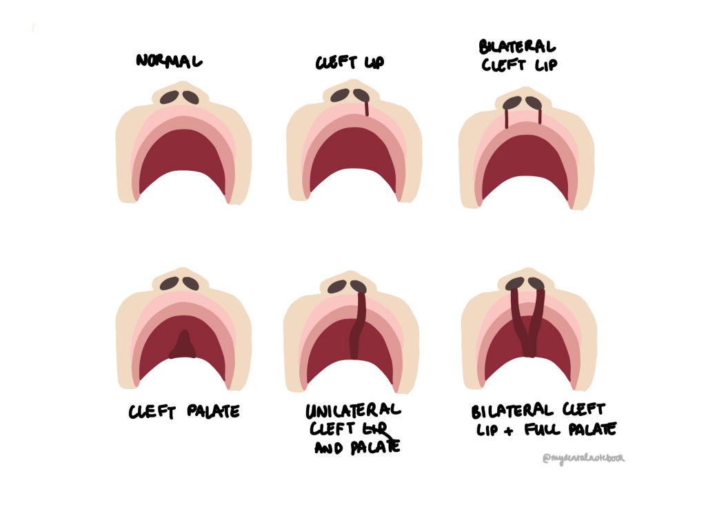
Over the next 1-2 weeks the shelves go through their fusion process, with themselves, the primary palate and the nasal septum. The epithelial surfaces from each shelf make contact forming a seam of desmosome. On contact communication pathways occur, causing cell death (apoptosis). This process if epithelial apoptosis allows the underlying ectomesenchyme to contact. The process begins centrally in the midline of what will be the hard palate. It continues anteriorly to then fuse with the primary palate as well as the inter maxillary segment of the face (lip and nasal fissures). From the midline it then travels posteriorly. The next phase of development will consist of the intramembranous ossification of the maxilla. The fusion process of the palate explains why so many cleft patterns can be seen clinically if the process was to be interrupted during development.

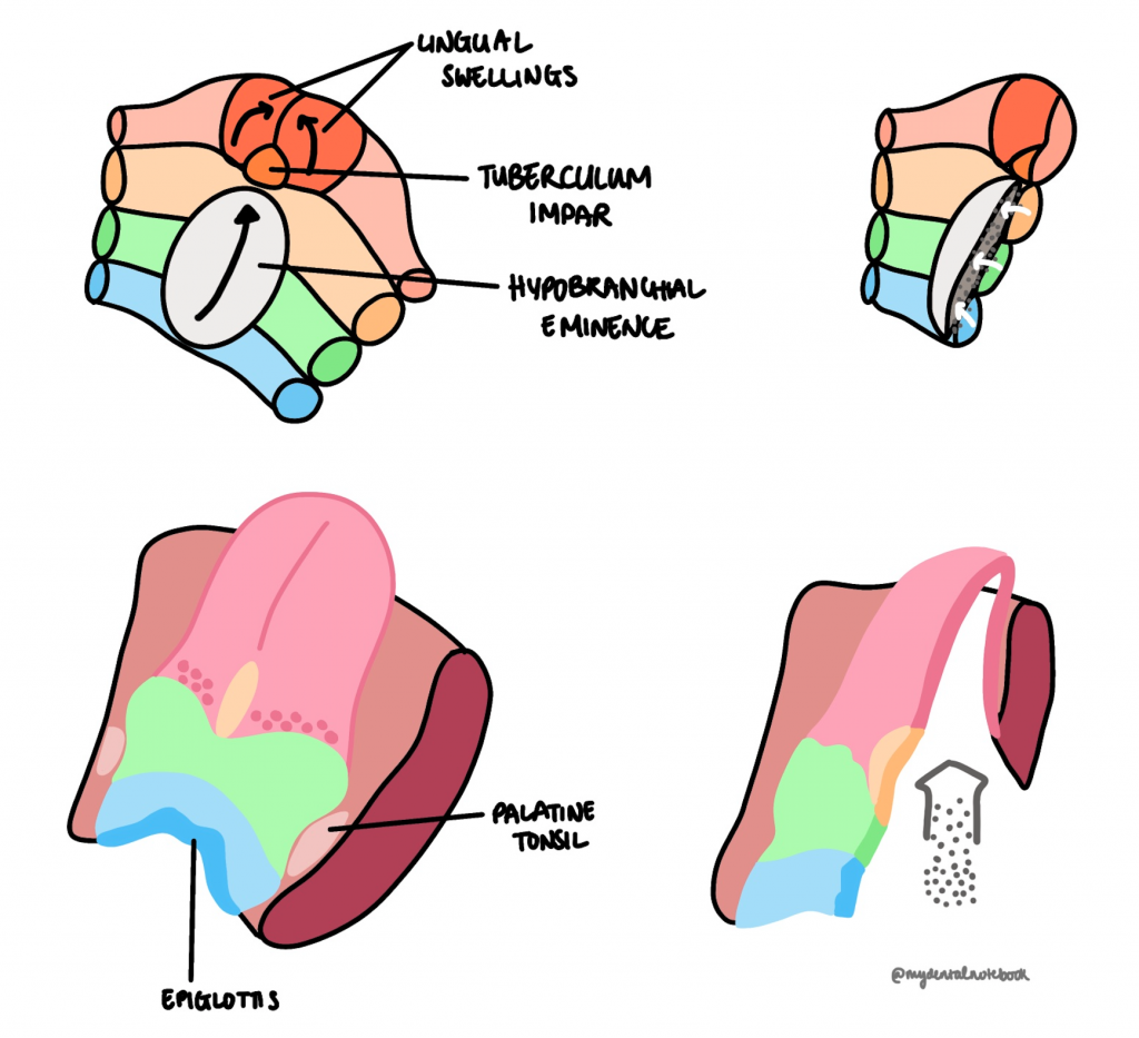
Understanding the embryology of the tongue helps to see why it has such a vast amount of different nerves innervating it. We can also see the clinical landmarks of the embryological development of the tongue: The lingual swellings that grow to form the anterior 2/3 give rise to the median sulcus which, we can see clinically at the midline. The sulcus terminals can also be seen clinically where arch three migrates forward to meet arch one. At the centre of this border there is the remanence of the foramen caecum, where the thyroid started its descent.
The skull consists of three main components: Cranial Vault, Cranial Base and the facial region. The vault forms via intramembranous ossification whereas the cranial base forms via endrochondrial ossification due to the cartilaginous precursor. We will look at the maxilla and mandible in a little more detail below:
The maxilla develops from one main centre of ossification, found around the area of where the canine would be situated. Intramembranous ossification continues as well as remodels. The maxilla when formed consists of its four processes: palatine, zygomatic, frontal, and alveolar. Remodelling is also required for the formation of the maxillary sinus.
The mandible initially forms via intramembranous ossification using Meckel's cartilage as a guide. Secondary sites appear as cartilaginous frameworks at the condyles, coronoid and symphysis; therefore, form via endochondrial ossification. There is no true articulating eminences at the TMJ of a new born; this forms around the same time the deciduous teeth begin to erupt. Meckel's cartilage goes on to form the sphenomandibular ligament (attaching at the lingual) as well as the malleus and incus (ossicles of the ear).
As the child grows and develops, further appositional (deposition and resorption) growth will occur alongside remodelling into the adult facial structure.

Around day 22 the lens placode will have developed and the beginning of the optic groove will appear. Through proliferation, differentiation and invagination of folds, the special layers of the eye will develop, producing the retina. For more depth of the development of the eye please use the reference to expand on your reading.
The otic placode forms and will go through many processes to produce the auditory pits and vesicles. Differentiation will occur to form the labyrinth and cochleae around 20 weeks. Remember pouch 1 will go to develop the eustation tube and cleft 1 forms the EAM and contributes to the tympanic membrane. Arches 1 and 2 will migrate and form what's known as the Hillocks of His and will eventually develop into the external ear pinna.
Cell migration and differentiation are fundamental during the embryological journey. As the pharyngeal arches come forward and the facial prominences develop, the framework is set up for the details to be created. Facial development is complex, but having the basics solidified really does help our understanding of the adult anatomy, for example the innervation of the muscles of mastication. I hope you found this lesson useful and enjoy the rest of the anatomy topic area.
General References used:
Berkovitz BK, Holland GR, Moxham BJ. Oral Anatomy, Histology and Embryology E-Book. Elsevier Health Sciences; 2017 Jul 11.
Nanci A. Ten Cate's Oral Histology-e-book: development, structure, and function. Elsevier Health Sciences; 2017 Aug 15.
Tortora GJ, Derrickson BH. Principles of anatomy and physiology. John Wiley & Sons; 2018 May 15.
Marieb EN, Keller SM. Essentials of Human Anatomy and Physiology: Books a la Carte Edition. Benjamin-Cummings; 2011.
Norton NS. Netter's head and neck anatomy for dentistry e-book. Elsevier Health Sciences; 2016 Sep 13.
Logan BM, Reynolds P, Rice S, Hutchings RT. McMinn's Color Atlas of Head and Neck Anatomy E-Book. Elsevier Health Sciences; 2016 Oct 21.
Atkinson ME. Anatomy for dental students. Oxford University Press; 2013 Mar 14.
Dental Update:
Alroyayamina S, McKnight M. Keep an eye out for the bifid uvula: a case report. Dental Update. 2015 Apr 2;42(3):247-9.
Dental Update: Part 1 of 3:
Part 1: McIntyre GT. Management of patients with non-syndromic clefts of the lip and/or palate part 1: from antenatal diagnosis to primary surgery. Dental Update. 2014 Oct 2;41(8):678-88.