This lesson covers the key life science principles of muscle structure and physiology. It is imperative to understand these concepts in order to apply these to the muscles of the head and neck and clinical practice.
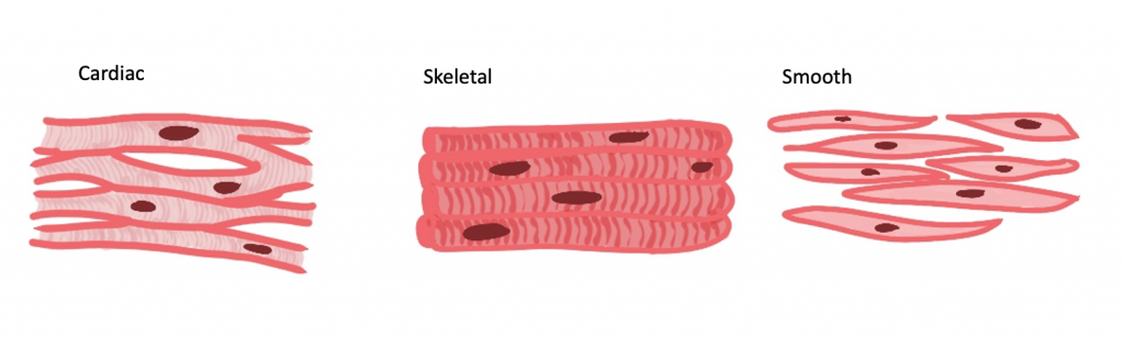
There are 3 types of muscles: skeletal, cardiac and smooth. Histologically, skeletal and cardiac muscle appear striated, whilst smooth muscle is non-striated.
The skeletal muscles are your voluntary muscles for movement and metabolic activity.
Whilst your cardiac muscle is self-explanatory - it can be referred to as your myocardium and is defined as involuntary movements as it is controlled by the autonomic nervous system.
Smooth muscle is commonly known to be found in blood vessels and controls vasoconstriction and vasodilation, but remember it can also be in the GI tract!
Skeletal muscles - which are the muscles involved in the head and neck region, attach to the bone via tendons (fibrous connective tissue).
If we look into a muscle step by step you will find that it is broken down into parts called fascicles. Each fascicle is made up by a number of 'muscle fibres' which are, in fact, cells. Each muscle fibre is made up of a number of myofibrils (organelle), which are a bundle of myofilaments (like a cytoskeleton).
As you can see from the description above, muscles are made up of many small units which work together to create muscle.
The myofibrils (small components of fascicles), have small areas along it called sarcomeres. These have the ability to expand and contract using a concept termed 'sliding filament theory'. These movements allow muscles to move as a result of nerve impulses. Keep reading for more information about how this works.
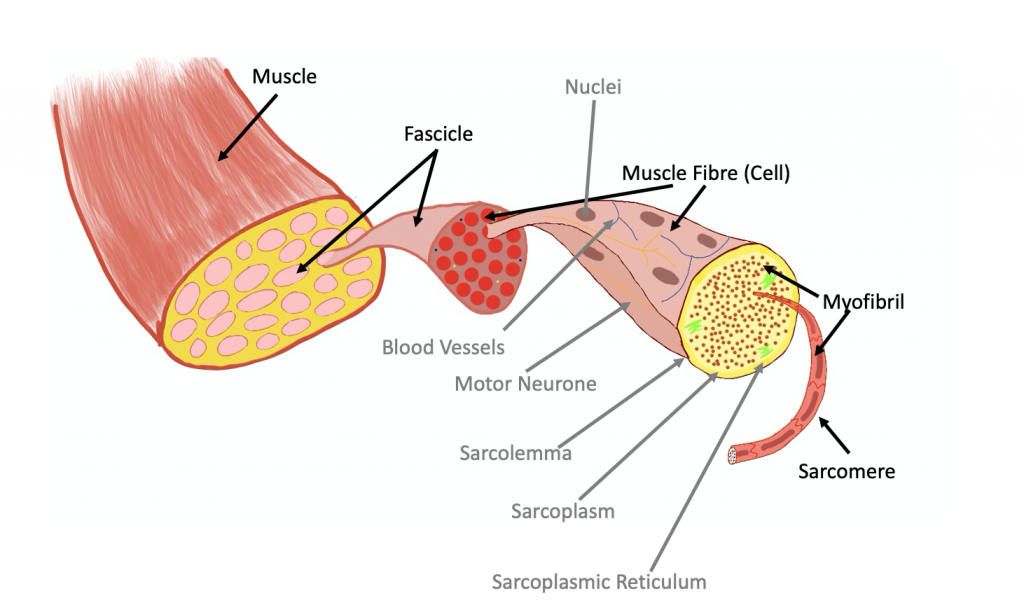
Each muscle fibre has a cell membrane; in muscles, this can be termed the 'sarcolemma'. The sarcolemma folds into the cell and forms 'T-tubules', whose function is to spread action potentials throughout the muscle cell.
The 'cytoplasm' of a muscle fibre can be termed the 'sarcoplasm'.
Remember, muscle cells are extremely active, so will have large stores of glycogen and plenty of mitochondria to produce ATP.
The myofibrils are surrounded by the sarcoplasmic reticulum (this is the muscle cell equivalent of the endoplasmic reticulum in normal cells, which has the primary function of protein synthesis).
Some example of structural myofilaments involved in the cytoskeleton in muscle cells are titin and dystrophin (1).
As mentioned above, myofibrils can be broken down into sarcomeres. These sarcomeres are demarcated by 'Z-lines'. They can be described as having light actin bands and darker myosin bands (note: actin and myosin are examples of myofilament proteins), which create the striated appearance.
The light actin bands are termed 'I bands' and sit on either side of the darker myosin 'A bands'.
The myosin filaments are connected together at an area termed the 'M line'.
There is a 'neutral zone' which contains no myofilaments, called the 'H band'. This is found in the centre of A band and allows space for the filaments to slide over one another.

As previously mentioned, Myosin and actin are myofilament proteins.
The thick myosin proteins have bulbous ends which are called 'heads'. The heads have 2 binding sites, one for actin and one for ATP.
The thinner myofilaments consist of actin, and two smaller proteins called tropomyosin and troponin.
Actin has 2 binding sites for the thicker myosin protein. When resting, tropomyosin spirals around the actin protein and covers these binding sites. Troponin binds the tropomyosin to the actin protein.

Muscles contract when the Z-lines pull the actin filaments inwards. These then begin to overlap the thicker myosin filaments.
Muscles contract when the actin filaments move towards the M-line (towards the middle). This means the actin filaments 'slide' past the myosin ones and hence this is termed the 'sliding filament theory'. The Z-lines therefore are pulled closer together and the sarcomere length shrinks.
In terms of bands, on contraction:

In order to contract, the myosin and actin filaments need to form a cross-bridge. This cannot occur at rest, as the tropomyosin blocks the myosin-binding sites on the actin filament.
However, when an action potential reaches a muscle cell, calcium ions are released by the sarcoplasmic reticulum in the sarcoplasm. These calcium ions bind to the troponin protein.
By binding to the troponin, it changes its structure and this pulls away the tropomyosin from covering the binding sites.
Now that the binding site is exposed, cross-bridge formation can happen. The following is more detail as to how this occurs.

This contraction process occurring in many small sarcomeres, causes the entire muscle to contract.
When the action potential reaches the terminal end of a motor neurone, it reaches a neuromuscular junction (in this case).

Muscle relaxation occurs when the calcium ions that were released by the sarcoplasmic reticulum are returned. The troponin molecule then returns to its normal structure and hence the tropomyosin sits over the myosin-binding sites on the actin filament.
As eluded to at the beginning of the lesson, smooth muscle is non-striated and involuntary movement.
It can be involved in digestion in the GI Tract, vasodilation and vasoconstriction in blood vessels and iris control in the eye, for example.
Smooth Muscle can be broken down into single-unit and multi-unit.
Single unit allows the muscle to contract, as a single-unit in one go, as a result of an action potential. This usually involves the action potential being dispersed quickly. This type of muscle can be found in the walls of visceral organs (excluding the heart).
Multi-unit smooth muscle does not allow an action potential to spread from one muscle cell to the next. These are regulated by the autonomic nervous system and some hormones. This type of smooth muscle can be found in the airway, and eyes for example.
Smooth muscle cells do not contain troponin and hence its contraction is controlled differently. The main protein in smooth muscle responsible for regulation of contraction is called calmodulin.
Calcium binds to calmodulin, which activates an enzyme called myosin-kinase. This phosphorylates the myosin heads - causing them to attach and pull the thin filaments. The entire muscle contracts in a corkscrew motion (1).
You should now be confident in understanding muscle structure and how muscles contract, particularly skeletal muscles as these are relevant to the head and neck.
Remember what a sarcomere consists of and which bands shorten in contraction, as this is a common exam question!
A handy tip - keep any old GCSE/A-level//IB/Notes from school for your Life-Sciences, they will help!