This lesson is made to provide the key elements to remember when learning the bones of the head and neck.


| Bone | Articulates | Landmarks | Foramina, boney pathways + Constituents |
Parietal | Frontal Parietal Temporal Occipital Sphenoid | superior and inferior temporal lines part of the temporal fossa parietal foramen meningeal grooves granula foveolae superior sagital groove | Parietal foramen: Emissary vein |
Frontal | Parietal Sphenoid Zygomatic Maxilla Ethmoid Nasal Lacrimal | Supra-orbital notch Glabella Frontal sinus Frontal crest superior sagittal groove menigmaal grooves Foramen caecum orbital roof | Foramen Caecum: emissary v. Supra-orbital notch: CN- V1 - Supra-orbital n |
Occipital | Parietal Temporal Sphenoid Atlas (C1) | Condyles Clivus - note: basilar part Sinus grooves: inf petrosal transverse occipital sup sagittal meningeal grooves internal and external protuberances Inf and Sup nuchal lines hypoglossal canal condylar canal external occipital crest | F. Magnum: Vertebral a. Meninges Medulla oblongata CN-XI - spinal root Hypoglossal canal: CN-XII - Hypoglassal Jugular foramen: (note: between the temporal and occipital bone) CN-IX - Glossopharyngeal CN-X - Vagus CN-XI - Accessory Sinuses: infer petrosal and sigmoid note: Internal jugular v. Posterior meningeal a. Mastoid foramen (note between temporal and occipital - inconstant) emissary v. and posterior meningeal a. Condylar canal: emissary v and meningeal b of pharyngeal a. |
Sphenoid | Frontal Parietal Occipital Temporal Maxilla Zygomatic Ethmoid Palatine Vomer | Wings: Lesser Greater Anterior clinoid process meningeal grooves Body Sella turcica Tuberculum Hypophyseal Dorsum posterior clinoid process Carotid groove Clivus (basilar part of occipital) Pterygoid plates: Later Medial Hamulus Spine Sphenoid sinus | Optic Canal: CN-II - Optic nerve Opthalmic a. Sympathetic plexus SOF: CN-III: oculomotor CN-V1: ophthalmic divi Lacrimal Frontal Nasocillary CN-IV: Trochlear CN-VI: Abducent Superior opthalmic v. IOF: (note: formed between sphenoid and zygomatic) Inf orbital v. Inf orbital a. CN-V2 - Inf orbital n. F. Spinosum Middle meningeal a. & v. F. Rotundum CN-V2 - Maxillary div F. Ovale CN-V3 - Mandibular div. (Note: lesser petrosal n.) F. Lacerum: (note: formed between the sphenoid and temporal bone) pathway of the - internal carotid a. Note: greater petrosal n. Sphenopalatine foramen: CN-V2 - nasopalatine and Posterior sup nasal |
Ethmoid | Inf cocha Frontal Sphenoid Palatine Maxilla Vomer Nasal Lacrimal | Orbital plate Perpendicular plate (bone part of nasal septum) nasal concha: Superior middle Crista galli Cribriform plate | Foramen: Ant ethmoidal: Ant Ethmoidal a/v CN-V1 - Ant Ethmoidal Post ethmoidal: Post Ethmoidal a/v CN-V1 - Post Ethmoidal Cribriform foramina: CN-I - Olfactory n. |
Lacrimal | Ethmoid Frontal Maxilla Inf concha | ||
Temporal | Parietal Occipital Zygomatic Sphenoid Mandible | Squamous part Petrous part Mastoid process Styloid process Zygomatic process Tympanic part - EAM - IAM Houses vestibular and Cochlear apparatus Mandibular fossa (Glenoid fossa) Articulating eminence Jugular fossa Grooves for the sigmoid and superior petrosal sinuses | Carotid canal: internal carotid a. Internal carotid autonomic nerve plexus Hiatus: lesser and greater petrosal n. IAM: CN-VII - Facial CN-VIII - Vestibular cochlear Labyrinth a. Canaliculus: Tympanic: CN-IX - Tympanic b. of glossopharyngeal n. Mastoid CN-X - Auricular b. of Vagus n. Stylomastoid foramen: CN-VII - Facial Petrotympanic fissure: CN-VII - Chorda tympani (intermediate b) Note: foramen lacerum (see sphenoid bone) Note: mastoid foramen (see occipital bone) |
Maxilla | Frontal Sphenoid Zygomatic Maxilla Ethmoid Palatine Vomer Nasal Lacrimal Inf Concha | Processes: Zygomatic Frontal Palatine Maxillary Alveolar Anterior nasal spine Infra-orbital foramen Orbital surface infra-orbital groove (note: IOF) fossa of the lacrimal sac Tuberosity Nasal surface Palatal surface (when together - inter maxillary suture) | Infra-orbital foramen CN-V2 - Infraorbital Alveolar foramina CN-V2 - Alveolar branches. Incisive canal CN - V2 - Nasopalatine Sphenopalatine vessels |
Palatine | Sphenoid Maxilla Ethmoid Vomer Palatine Inf concha | Pyramidal process Orbital process Horizontal plate Perpendicular plate Posterior nasal spine Greater and lesser palatine foramina | Foramina: Greater: CN-V2 - greater palatine nerve Lesser: CN-V2 - lesser palatine nerve Note: also vessels |
Nasal | Nasal Frontal Maxilla | ||
Vomer | Sphenoid Palatine Maxilla Ethmoid | ||
Inf concha | Ethmoid Maxilla Palatine Lacrimal | ||
Zygomatic | Maxilla Frontal Temporal | Processess: Frontal Temporal Orbital surface | Zygomaticofacial foramen CN-V2 - Zygomaticofacial |
Mandible | Temporal (TMJ) | Body: Regions: Base Alveolar mental protuberance Mental formanen Mylohyoid ridge/ line Submandibular fossa Sublingual fossa Ramus: Processes: Coronoid Condylar Angle Mandibular notch Mandibular foramen Lingula Mylohyoid groove (begins) | Mental foramen: CN-V3 - Mental n Mandibular foramen: CN-V3 - Inferior alveolar Mylohyoid groove: CN-V3 - Mylohyoid n and vessel pathway |
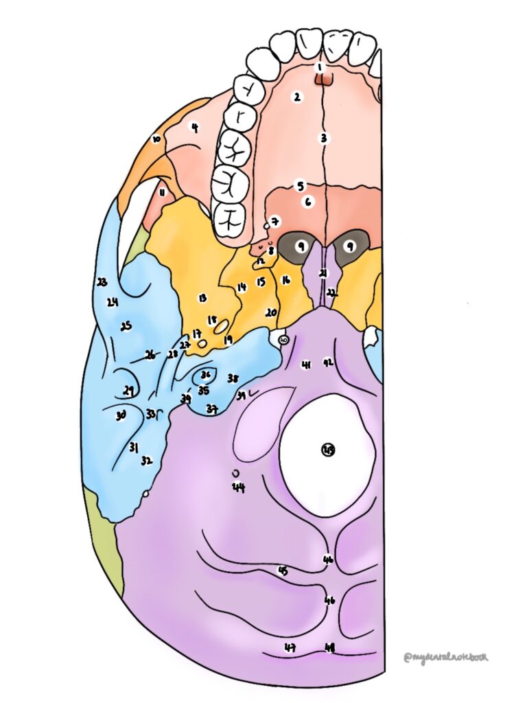


One of the most complex anatomical areas of the head and neck. Contains:


This lesson provides the "footnotes" you'll need when revising the bony anatomy or the head and neck. I would advise to play with a skull, take it apart and really get a feel for its orientation. There are endless books out there and some helpful ones are listed below - see references and third party reading.
General Referencing and further reading:
Atkinson ME. Anatomy for dental students. Oxford University Press; 2013 Mar 14.
Arx TV, Lozanoff S. Clinical Oral Anatomy.
Logan BM, Reynolds P, Rice S, Hutchings RT. McMinn's Color Atlas of Head and Neck Anatomy E-Book. Elsevier Health Sciences; 2016 Oct 21.
Norton NS. Netter's head and neck anatomy for dentistry e-book. Elsevier Health Sciences; 2016 Sep 13.