NOTE – Please note this assessment and management criteria are indicated by the British Society of Periodontology. If you are following this lesson from another region, please check your national guidelines in regard to periodontal assessment and management.
| Supracrestal attached tissues (previously termed Biological width) | The distance from the base of the gingival sulcus, to the alveolar bone; it includes the connective tissue and the junctional epithelium attachment to the tooth. |
| Calculus | Plaque which has calcified due to mineral deposits such as calcium and phosphates from the saliva |
| Furcation | The area where roots separate; this can be in a bifurcation (two roots), or trifurcation (three roots) |
| Mobility | The displacement of a tooth beyond its normal physiological boundaries in a horizontal or vertical plane |
| Periodontal Pocket | A pathologically deepened gingival sulcus |
| Pocket Depth | Is the distance between the free gingival margin and the bottom of the pocket. |
| Clinical Attachment Loss (CAL) | CAL is the distance between the CEJ and the bottom of the pocket. |
A BPE probe is the standard probe used in every initial dental assessment. This probe is used to give a BPE score.
Its structure has a 0.5mm ball at the end; this is ball shaped to prevent trauma to the gingivae, but also this will pick up the tactile feel of calculus subgingivally.
There are 4 sections to a BPE probe, with 2 black bands. The first black band is 3.5mm - 5.5mm. The second black band is from 8.5mm – 11.5mm.

A Williams probe can be used in a 6PPC to take more thorough and accurate measurements of the pocket, and the gingival recession. The Williams probe has markings at; 1, 2, 3, 5, 7, 8, 9, 10

A UNC15 probe can be used in a 6PPC also to take more thorough and accurate measurements of the pocket, and the gingival recession. This probe has black markings every 5mm. Therefore 5, 10 and 15! The UNC15 probe has markings at; 1, 2, 3, 4, 5, 6, 7, 8, 9, 10, 11, 12, 13, 14, 15

A Nabers probe is a probe which is used to measure the extent and the depth of a furcation. A furcation is the area where tooth roots meet, so can only be used in molar teeth. The markings on a Nabers probe are; 3-6mm, 9-12mm both indicated through a black band

The periodontal exam is divided into multiple stages depending on findings. There are multiple parts to be considered;
If a person shows to have signs of periodontal disease, a further assessment is used called a 6-point pocket chart (6PPC). This is divided into several further stages.
This lesson will guide you through the way to carry out a periodontal assessment to gain the appropriate information to make a diagnosis.
When initially assessing a patient, it is important to not only take measurements, but to also evaluate clinically the health of gums.
Clinical Health can be determined by;
Following a clinical inspection of the gums to give you an idea of the health, it should be followed by a Basic Periodontal Exam (BPE) as indicated by the British Society of Periodontology (BSP).

The BPE is the screening process for gum disease. This means exactly what it says in the name; basic. It allows dentist to check in the least invasive and discomforting manner if there is gum disease present, and also acts as a guide on where improved oral hygiene may be indicated.
A BPE is conducted using a BPE probe (sounds logical?). The BPE probe is walked around all 6 surfaces of the teeth with a light probing force estimated to be 20-25g. The clinician will be watching the depth the probe goes to and always watching the black band, as well as gaining information through tactile feedback for calculus or overhangs.
In a BPE, the teeth are sectioned into sextants. All the teeth in that sextant, excluding wisdom teeth, are included. The highest score is then taken as the recording for the sextant.
| Disto-Lingual | Lingual | Mesio-Lingual |
| Disto-Buccally | Buccally | Mesio-Buccal |
| 7,6,5,4 | 3,2,1,1,2,3 | 4,5,6,7 |
| 7,6,5,4 | 3,2,1,1,2,3 | 4,5,6,7 |
A full BPE should be conducted on all patients over the age of 18. A simple BPE should be conducted on patients over the age of 7, when they have their first permanent molars, and their permanent central incisors. More information regarding this can be found later in the lesson. The meaning of the band height, and the BPE it is therefore scored can be found below.
| Band | Pocket Measurement | BPE score | Meaning | |
| First black band completely visible | 0 - 3.5mm | 0 | No gingival bleeding No calculus or plaque retentive factors | |
| First black band completely visible | 0 - 3.5mm | 1 | Gingival bleeding No calculus or plaque retentive factors | |
| First black band completely visible | 0 – 3.5mm | 2 | Presence of calculus or/and plaque retentive factors | |
| First black band is partially visible | 3.5mm - 5.5mm | 3 | Indicates a pocket depth between 4-5mm | |
| First black band is entirely in the pocket | >5.5mm | 4 | Indicates a pocket depth above 6mm | |
| - | - | * | This means a furcation can be felt. This is added onto the BPE score eg 4* |

Therefore, the BPE for this patient would be:
| 4* | 1 | 3 |
| X | 2 | 3 |
| BPE | Management | Additional Investigations |
| 0 | No further guidance required | No investigations required. Reassess BPE at next checkup. |
| 1 | Oral Hygiene Instruction | Plaque and Bleeding Indices. Reassess BPE at next checkup. |
| 2 | Oral Hygiene Instruction, and removal of the plaque retentive factors eg. Supra/Subgingival calculus | Plaque and Bleeding indices. Reassess BPE at next checkup. |
| 3 | Oral Hygiene Instruction, removal of the plaque retentive factors, and Root Surface Debridement (RSD) if necessary | Plaque and Bleeding indices. Radiographs should be considered. 6PPC indicated in only the sextants of 3 after initial therapy. |
| 4 | Oral Hygiene Instruction, and RSD. Consider referral to a specialist for more complex treatment. | Plaque and Bleeding indices. Radiographs taken to indicate attachment loss. 6PPC before and after treatment. |
| * | Treat based on the BPE code. Consider referral to a specialist for more complex treatment. | 6PPC before and after treatment. |
The BPE on a child can feel strange to do, especially as they are so young that having a probe in their gums is going to feel very weird and can be uncomfortable. This means that an alteration to the BPE is used, termed the simple BPE, until they are older and therefore more accustomed to the gum exams a dentist might do! A Simple BPE should be conducted on patients over the age of 7 when their first permanent molars, and permanent central incisors have erupted.
It is especially important to recognise the scoring and the teeth noted as it is very common for false pocketing during the mixed dentition phase, which can falsely indicate the need for treatment.
Bleeding on probing (BoP) helps to give dentists an idea of the gum health, as inflamed gums will bleed. This means that active disease is occurring.
During the process of periodontal treatment, it is very important to repeat these bleeding indices as it gives the patient a goal.
For example, in their initial appt they have a score of 50%, but in their second appointment after meticulous oral hygiene instruction it is 35%. This allows the patient to see their efforts to help improve their oral health. It also can indicate where more help is required.
If a patients BoP scores stay high, it means that there is active periodontal disease still occurring indicating poor patient compliance or difficulty; this can influence the prognosis for future restorative and prosthodontic treatment.

A Plaque index is a very helpful diagnostic and visual aid to help a patient understand what you are looking for! It works to test the amount of plaque present on the teeth. It typically involves disclosing the plaque using food colouring which adheres to bacterial biofilms therefore highlighting their presence.
Prior to beginning treatment, a plaque index should be taken. This allows the patient to understand their oral health status to act as motivation for improvement, very similar to their bleeding index. Throughout the treatment, these indices should be repeated, especially prior to carrying out prosthodontic treatment as it is likely to have a poor prognosis if plaque levels are not controlled.
There are several types of plaque indexes which can be used as described below;
This method measures the plaque accumulation on certain teeth
This method measures the amount of plaque present on each tooth.
This method measures the presence or absence of plaque on each surface of each tooth
Note: Prior to staining a patient’s teeth, ensure you offer them Vaseline to put on their lips. This will prevent the staining from transferring to their lips and giving them an interesting lipstick!

Radiographs are used in dentistry to support a clinical observation. They are also highly important at helping to determine the prognosis of teeth.
Radiographs are typically indicated after a clinical assessment, to either aid diagnosis or aid further investigation. With relevance to periodontal assessments, radiographs can give valuable information about the extent (generalised or localised), severity (mild, moderate, severe) and the pattern (horizontal, vertical, furcation involvement bone loss) of bone loss. These are usually taken when patients exhibit a BPE of 3, 4 or * or there is a clear clinical evidence of interdental recession
Horizontal bitewings can be taken however in cases of severe bone loss, these are not as helpful. Especially as they do not show additional pathoses which can be present in periodontally-involved teeth; consequently, affecting their prognosis. Vertical bitewings usually show more of the bone crest of periodontally involved teeth but still they cannot be used to assess the periapical status and the length of the root of the exposed teeth.

The gold standard for a periodontal assessment is a periapical (PA) radiograph. This is able to give a full picture of the tooth; including assessing the association of the level of the bone crest relative to the length of the root, furcation involvement, periapical and endodontic status of the teeth, and bony defects – all of which need to be taken into account to determine prognosis at tooth and patient level. In generalised periodontal disease, it is typically appropriate for full mouth PA's to be taken to aid the diagnosis.

OPGs can be helpful as they produce an overall picture of the dentition, including the height of the bone. However, they lack the detail and resolution for some teeth. Consequently, they may need to be supplemented with additional periapical radiographs, especially in the anterior sextants, due to superimposition of the image.

These 6 positions are checked for every tooth in the sextant after initial therapy if there is a BPE of 3, however all teeth in the mouth are checked if there is a BPE of 4.
During a 6PPC, much more information regarding the periodontal health is noted.
| Disto-Lingual | Lingual | Mesio-Lingual |
| Disto-Buccally | Buccally | Mesio-Buccal |
Above are the 6 positions which must be recorded for pocket depth and recession during a 6PPC.
Note - For general dental practitioners, it is the minimum requirement to record pocket depths and BoP in a 6PPC however this will not give you as much clinical data therefore the recommendation is to complete all sections!
CAL is a more accurate indicator of the periodontal destruction around a tooth than probing depth alone. CAL defined as the distance between the CEJ and the bottom of the pocket. It aims to calculate the degree of periodontal disease taking into account active disease (pocket depth) and evidence of previous disease as shown through recession.
Where recession has occurred, CAL can be calculated as follows;
Pocket depth + Recession = Clinical Attachment Loss
In cases of gingival enlargement, to calculate the CAL we need to subtract the distance between the gingival margin to the CEJ from the pocket depth (PD)
CAL = Pocket depth - distance from gingival margin to CEJ

Recession of the gums is when the gums have started to cover less of the teeth; this can include the loss of interdental papilla. There are a number of causes for recession including abrasive tooth brushing, periodontal disease, periodontal therapy such as scaling, tooth positioning or a combination of the above.
Patients showing recession will report their teeth look longer or are more sensitive. Therefore, understanding the cause for the recession is essential to prevent its progression. Below are some examples where recession can be avoided or indicators of another problem:
If there is a history of periodontal disease, unfortunately recession is unavoidable. During periodontal therapy, the inflamed gum will recede, therefore reducing the pocket depth. This is a positive outcome as the pocket becomes easier to clean, despite the gum recession.
Once gums have receded, the tooth is more subject to problems. For example, root caries, dentine hypersensitivity, and poor aesthetics. Communicating this information to patients is very important prior to carrying out non-surgical/surgical periodontal treatment. This means that the patient isn't surprised if afterwards their gums recede.
Remember – it's an excuse if you tell them after!

Furcation involvement occurs when the bone loss has extended to the level of where the roots separate in molars; this can be in a bifurcation (two roots), or trifurcation (three roots). Grading a furcation focuses on the horizontal measurements rather than the vertical, meaning a specialised curved probe, called a Nabers probe is used to measure the amount of bone loss in the horizontal plane.
| Clinical Observation | Grade | Representation of Probe through Furcation |
| No furcation present | 0 | - |
| Probe passes into the furcation however does not extend more than 1/3rd of the bucco-lingual width of the tooth | 1 |  |
| Probe passes into the furcation however does extend more than 1/3rd of the width of the tooth, however does not extend completely through the furcation | 2 | 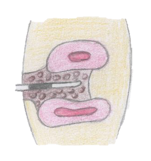 |
| Probe extends completely through the furcation | 3 |  |
Furcation involvement must be noted as it can affect the prognosis for the tooth affected. Especially as keeping this area clean is very difficult for the patient. However, various surgical and non-surgical techniques can be used to ease the cleaning and improve the prognosis.
Tooth mobility is the amount a tooth can move. Teeth naturally have the ability to move within their socket a minimal amount of around 0.2mm horizontally; this is facilitated by the PDL. However, it is very common in the later stages of periodontal disease for a patient to become more aware of tooth movement or 'looseness'.
The way to think about it, is like you have a post in the ground, surrounded by soil. If you dig up some of the soil, it allows the post to wobble a bit more as it's not as well supported. However, if you keep digging up the soil around the post, eventually the post will fall over.
Applying this to dental terms, the tooth is the post and the bone is the soil. Periodontal disease leads to the loss of bone, meaning the tooth can move more, and can eventually fall out. It can also occur if the tooth is subject to excessive occlusal loading.
| Clinical Observation | Score |
| Mobility is under 0.2mm (this is a healthy tooth's physiological mobility) | 0 |
| Mobility is between 0.2mm - 1mm in the horizontal plane | 1 |
| Mobility is over 1mm in the horizontal plane | 2 |
| Mobility is in both the horizontal and vertical plane | 3 |
NOTE: this was previously termed Biological Width however has now been replaced by the term Supracrestal Attached Tissue. (1)
The supracrestal tissue attachment is a measurement which describes the distance from the base of the gingival sulcus, to the alveolar bone. This is said to be approximately 2.04mm but it varies amongst individuals (2). The importance of the supracrestal tissue attachment comes in when we perform restorative work such as crown and bridgework, for example. The margins of our restorations should be maintained ideally supragingival or at the same level of the gingival margin. Sometimes it may be necessary to finish our preparations slightly subgingivally. This poses the risk of violating the supracrestal tissue attachment (or more commonly known to clinicians, biological width, although this term has now been replaced). An infringement of restorative margins within the supracrestal connective-tissue attachment is associated with inflammation and loss of periodontal supporting tissue.
The tissues may try to re-establish the desired amount between the margin of the restoration and the bone crest either by a pocket formation and bone loss or by gingival recession. This is considered to be dependent on the gingival biotype of the patient (thick or thin respectively).
Therefore, if there is an area of localised gingival recession, or bone loss with generalised good oral hygiene, always consider if this is related to an overextended restoration which impinges on supracrestal attached tissues. This means that the gingival sulcus and bone will adapt to maintain a constant width. In the planning stages for restorative treatment this must be a consideration to prevent avoidable damage.

A basic periodontal assessment should always include information collected through;
For a more detailed periodontal assessment, a 6PPC should be used to gain more information.
Specific References:
(1) Caton JG, Armitage G, Berglundh T, Chapple IL, Jepsen S, Kornman KS, Mealey BL, Papapanou PN, Sanz M, Tonetti MS. A new classification scheme for periodontal and peri‐implant diseases and conditions–Introduction and key changes from the 1999 classification. Journal of periodontology. 2018 Jun;89:S1-8.
(2) Gargiulo AW, Wentz FM, Orban B. Dimensions and relations of the dentogingival junction in humans. The Journal of Periodontology. 1961 Jul;32(3):261-7.
General References:
Bosshardt DD. The periodontal pocket: pathogenesis, histopathology and consequences. Periodontology 2000. 2018 Feb;76(1):43-50.
British Society of Periodontology. Young Practitioners Guide to Periodontology. British Society of Periodontology; 2012.
Clerehugh V, Tugnait A, Genco RJ. Periodontology at a Glance. John Wiley & Sons; 2013 May 31.
Ziada H, Irwin C, Mullally B, Allen E, Byrne PJ. Periodontics: 1. Identification and diagnosis of periodontal diseases in general dental practice. Dental update. 2007 May 2;34(4):208-17.