In this lesson we will look at the special investigations that should be considered prior to extractions. The first part of the lesson provides an overview of many of the prognostic investigations that should be carried out; to read in more depth regarding these, please refer to the Endodontic and Periodontal areas of the site. This lesson will focus mainly on the general radiographic assessment of the tooth to be extracted. Specific teeth e.g. third molars, will be covered in future lessons.
This is the pulps ability to respond to stimulus [1].
Electric pulp test (EPT), contains two parts one metal hook to be held by the patient or placed in the buccal vestibule, and a probe which is placed on the tooth. (The patient can pull apart the device if it is to be held, allowing them control to stop the stimulus).
The EPT probe needs be dipped in toothpaste to be effective. It has been suggested that placing the probe tip within the incisal/occlusal two thirds of the crown provides more consistent results [1].
EPT can have false readings in some instances, e.g. pulp calcifications can insulate the current.
Using a cotton wool pledget with a cold spray such as ethyl chloride is more reliable in gaging pulpal health. Other superior variants include Dichlorofluoromethane and CO2 snow [1]. The cold test has been shown to be more reliable than EPT [1].
Yes the dreaded 6 point pocket chart can be useful in diagnosing the tooth!
This is very useful especially around tooth in question, as a very deep localised pocket can be consistent with vertical root fracture. Probing the tooth will allow you to gage bone loss and periodontal health. Any active bleeding is a sign of active periodontal disease and any pus exudate from the pocket is a sign of infection and should be recorded in the notes.
This can help gage the extent of periodontal disease; the greater the furcation the more advanced the periodontal disease.
This will help gage the difficulty of extraction, as pre-operatively mobile teeth should be easier to extract. Note: in the older patient, the tooth may be highly restored and sat in brittle not very elastic bone; therefore, could be more likely to fracture. ("beware the lone standing molar")
Any pain or tenderness during percussion testing or applying apical or lateral pressure to the tooth should warrant further investigation as this is consistent with a peri-radicular infection. Tenderness on palpation of the surrounding periodontium could also indicate this (note: any swelling?)
Cracked tooth syndrome (CTS) can be the reason behind the pain and can be tricky to differentiated from periapical periodontitis. However, pain on releasing the bite ("rebound pain") can be a differentiating factor. Note: Pain on biting ("application pain") does commonly occur. CTS is when a tooth has an incomplete fracture/ "hairline" (cusp still attached to tooth) of the enamel and dentine commonly found in a vital posterior tooth. The crack may extend into the pulp. The teeth most commonly affected by CTS are Lower 7s, 6s and upper premolars [2].
The pain can be tested by replication. Using a cotton wool roll for the patient to bite on or a tooth slooth [2]. The tooth slooth, also known as fractfinder is a more accurate method of detecting fractures [2]. The tooth slooth can be placed on each individual cusp and the patient is asked to bite allowing selective pressure on each individual cusp. Pain on biting or release is indicative that cusp is fractured [2]. Note: A vertical root fracture is usually a terminal prognosis.
Diagnosing pulpal and peri-apical conditions are explored in more depth under the Endodontic category: Diagnosis of pulpal and periapical diseases.
Following on from previous lessons, you may be familiar with the contents of the WHO form which helps reduce the complications of surgery [5]. It is a pre-requisite to dental extractions to have the pre-operative radiograph available for viewing peri-operatively, as it helps the clinician assess the difficulty of extraction(s). For the purpose of this lesson we shall consider the radiograph is available in front of us, and will discuss different scenarios. The radiograph should ideally be a periapical radiograph to allow full visualisation of the tooth in question and be adequate for the surgical assessment[3, 4]. You may see on placements an OPG and other image types e.g CBCT being used.
We will now look at some of these factors in more detail below:
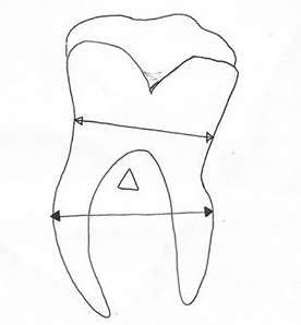
If the width of the roots is wider than the crown this may require a surgical approach to section the roots [4]. The roots are measured at their widest point (white arrow) and compared to the widest portion of the crown (black arrow at contact points), if the root measurements are wider than the contact point this can indicate an inadequate path of withdrawal (Upwards arrow) therefore, may require a surgical approach [4].
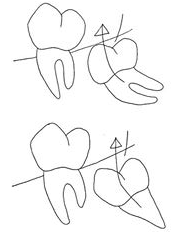
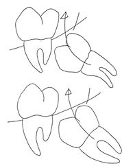
Angulation of the crowns and roots. Note: Dilacerations are sharp bends between the crown and root, resulting in an inadequate path of withdrawal [4].
Visualise the total number of roots is sometimes difficult as the roots are super imposed and appear as a faint shadow radiographically [5]. The number of roots will influence the extraction technique e.g. a figure of 8 motion when extracting.
In summary; the longer, thinner and more curved the roots the harder the extraction due to an increased risk of root fracture [4].
Radiolucency’s in pulp space/ canal are a sign of internal resorption and can complicate extractions as the affected area is more fragile, carrying an increased risk of fracture.
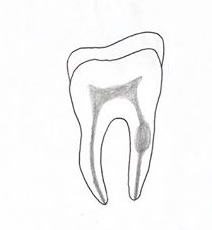
Note: Radio-opaque pulp space/ canal is consistent with root treated teeth (e.g. GP filling material) and care should be taken during extraction as root treated teeth can be brittle.
The surgical sieve for this includes a lot of conditions; however, for the context of XLA radiographic assessment, we are considering:
Hypercementosis and condensing osteitis appear as a round radio-opaque mass around the roots. To decipher the difference between them look at the PDL space. Hypercementosis appears as a radio-opacity contained within the PDL, whilst condensing osteitis is confined to outside the PDL space [3].
Ankylosis of roots can also be detected by percussion test. Commonly ankylosed teeth make a more metallic sound. Radiographic appearance of ankyloses is characterised by absence of the PDL space.
Extracting teeth requires a degree of expansion in the bone [4]. If the supporting bone is thick or dense, achieving adequate expansion can prove to be more difficult, putting the roots at risk of fracturing. Patients with parafunctional habits (grinding teeth) commonly have thick and dense alveolar bone. Note: buccal exostosis also makes bone expansion more difficult. In these circumstances a surgical approach may be appropriate to reduce the risk of the tooth or buccal plate fracturing [4].
Proximity to the surrounding structures such as the inferior alveolar nerve, mental nerve or maxillary sinus should be considered radiographically, as this will determine the risk associated with the extraction and whether a surgical approach should be a considered alternatively [6].
Communication between the mouth and the maxillary sinus can occur following the removal of upper molars and premolars. This communication is called an Oro-antral communication (OAC) [7]. Patients should be warned that this communication between the mouth and sinus may require further surgery to close. If left untreated it can go on to form an Oro-antral fistula (OAF).
Molar and premolar teeth risk causing an OAC particularly if [4]:
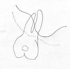
Lower third molars are associated with inferior alveolar and lingual nerve damage. This can be temporary or permanent. The incidence has been reported as high as 15% post-operatively and permanently as high as 0.3-0.6% [7].
Radiographic risk factors for IAN injury include [8]:
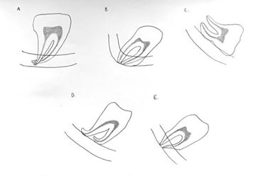
Note: Additional investigations such as Cone Beam Computerised Tomography (CBCT) can provide a 3D image, this can be especially helpful if some of the above radiographic risk factors are identified [8].
The incidence of tuberosity fractures as a complication of upper molar extractions is relatively low. A study comparing the prevalence of complications in 8455 routine extractions reported the rate of tuberosity fractures to be as low as 0.15% [9]. Fracturing the maxillary tuberosity can carry a bleeding risk [10] with the vascular supply deriving mainly from the maxillary artery and other fragile vessels. Preserving the tuberosity is also advantageous when considering future support and retention for upper dentures.
This lesson has provided a broad overview of the investigations needed before taking a tooth out. Please refer to the third party area and references to take your reading further.
A clinical guide to oral surgery book 1 - Tara Renton